The RTVue is the next generation 3D Optical CoherenceTomography sytem developed by the original inventors of OCT and the people who developed the first OCT system of eye-care 3D OCT is a non-invasive, noncontact, transpupillary Imaging technology, with ultra-high speed 26,000. A-Scan/second and high resolution 5 Microns Fourier domain OCT technology allows the systems advanced clinical protocols to be used for ocular examination with unprecedented resolution and clarity. Cross-sectional images of the retina are produced using the optical backscattering of light in a fashion analogous to B-scan ultrasonography. The anatomic layers within the retina can be differentiated and the retinal thickness can be measured more precisely than the conventional OCT. Besides, the optic disc and nerve fibre layer can be assessed in cases of glaucoma.The system also provides many advanced imaging features for comprehensive 3-Dimesional Retina and Glaucoma analysis
CIRRUS HD OCT 5000
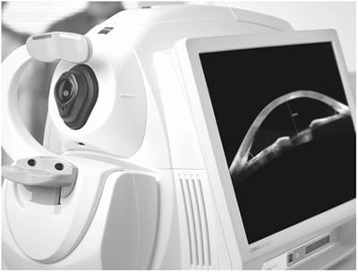 The next generation 3D Optical Coherence Tomography system.It is a noninvasive, non contact, transpupillary Imaging technology, with ultra-high speed 27,000 axial scans per second which allows 50 times faster data acquisition in practice and high resolution 5-µm axial and 15-µm transverse resolution in tissue . Fourier domain OCT technology allows the systems advanced clinical protocols to be used for ocular examination with greater resolution and clarity. The OCT uses light waves to make a map of retina, anterior chamber, cornea to show up any damaged areas. It uses optical principle known as low coherence interferometry to scan across the targeted eye structure and generate an image of the same. The procedure requires you to sit in front of the OCT machine and place your chin on chin rest and forehead touching the head support. You are required to focus at a given target and keep your eye still while the scan is being performed. Your scan will be evaluated by our doctors and discussed with you.
The layers within the retina can be differentiated and the retinal thickness can be measured. Also, the change from previous visit, effect of treatment can be analyzed.
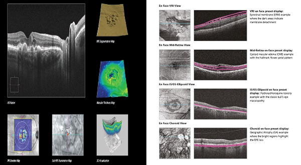
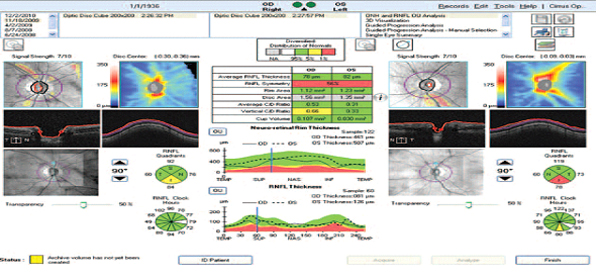
The optic disc and nerve fibre layer can be assessed and guided progression analysis provides valuable information in management of Glaucoma.
Anterior segment imaging of the angle and cornea and measurement of central cornea thickness can be done.
Forum Viewing System
This system is used to integrate scans of fundus, fundus fluorescein angiography and OCT which can be accessed by multiple devices for comprehensive analysis.
When to see a doctor
Make an appointment for an eye exam if you notice any changes in your vision. If you develop sudden vision changes, such as double vision or flashes of light, sudden eye pain, or sudden headache, see your doctor right away.
Fundus Fluorescien Angiography (FFA)
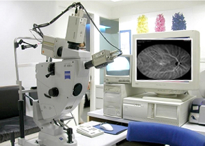
 This investigative procedure comprises of injecting a dye -FLUORESCEIN into one of the veins in your arm and either taking rapid serial photographs of its passage within the delicate blood vessels of the eye in the retina and choroid , using a digital Fundus camera or HRA or rarely examining the inside of your eye with an INDIRECT OPHTHALMOSCOPE using appropriate filters.
The information obtained from this test aids your doctor in making a diagnosis and planning treatment especially using different kinds of laser such as Diode, Argon,532 DF YAG PDT or TTT.
Apart from the needle prick and the light flash of the camera there is no discomfort from this test. If nausea appears remain calm and take deep breaths which helps in overcoming it. Your usual diet can be taken soon after the procedure. Fluorescein is highly non-toxic drug. In rare Instances, it can produce allergic reaction, which responds rapidly to appropriate medication.Serious life threatening allergic reactions, though exceptionally rare, can occur. The skin and urine may appear yellow in colour for about 36 hours following the test,and is not a cause of any concern .
Instructions To Patients:
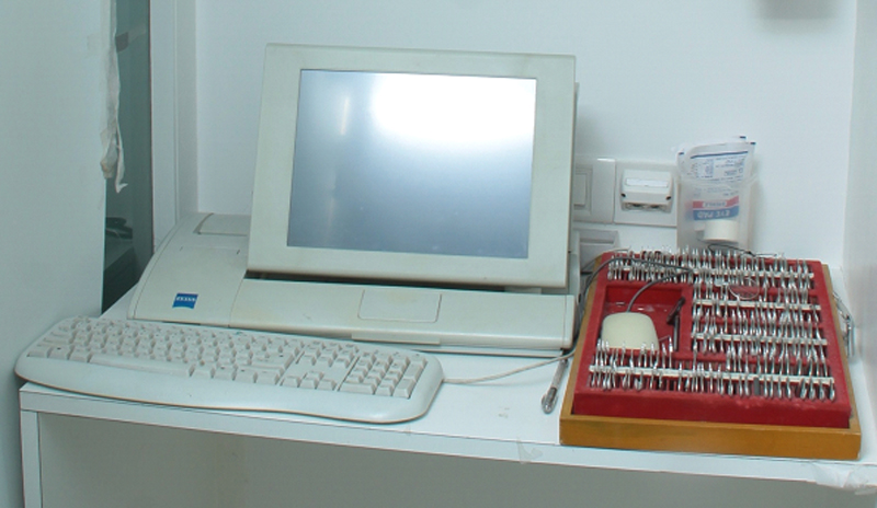
Equipments:
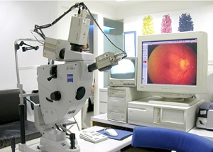
At the core of every successful treatment is a reliable diagnosis. This is why ZEISS has combined the diverse functions of sound retinal diagnostic procedures to produce one pioneering solution: the FF450plus fundus camera. In combination with the VISUPAC® image processing system, it allows for a unique level of diagnostic accuracy. It delivers images with the clearest details, thus providing the dependable basis for first-class treatment outcomes.
The renowned optical quality of the FF450plus and integration with VISUPAC’s digital imaging capabilities makes the difference. The result is images of exquisite detail – a must for today's demanding ophthalmic professionals – be it for disease documentation, presentations, or for use in clinical trials. Its optical zoom features three magnification levels (20°, 30° and 50°) for optimal diagnostic insight. In addition, the highly flexible tilt and swivel mechanism allows imaging from virtually any angle.
The exclusive ZEISS telecentric optics of the FF450plus provide constant magnification, independent of focusing and position, resulting in virtually no distortion. Together with VISUPAC’s uniquely harmonized sensors, this enables you to accurately and repeatedly determine and analyze disease progression.
Indocyanine Green Angiography (ICGA)
 This investigative procedure comprises of injecting a dye -INDOCYANINE GREEN into one of the veins in your arm and either taking rapid serial photographs of its passage within delicate blood vessels of the eye in the retina and choroid, using a digital Fundus camera of HRA using appropriate filters.
The information obtained from this test aids your doctor in making a diagnosis and planning treatment especially using different kinds of laser such as Diode,Argon,532 DF YAG, PDT , TTT or ICG enhanced photocoagulation.
Apart from the needle prick and the light flash of the camera there is no discomfort from this test.If nausea appears remain calm and take deep breaths which helps in overcoming it.Your usual diet can be taken soon after the procedure. Indocyanine Green is highly non-toxic drug.In rare Instances, it can produce an allergic reaction, which responds rapidly to appropriate medication, Serious life threatening allergic reactions,though exceptionally rare, can occur.
Instructions To Patients:
Anterior Segment Diagnostics
Tomey TMS-4 tomographer
It is Used for following purposes-
Oculus Pentacam
Zeiss IOL master
For measuring axial length of eyeball, corneal curvature and calculating IOL power before cataract surgery.
Ultrasonic A scanner and biometer Tomey UD 6000
For calculating axial length of eye.
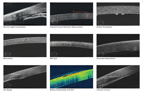
Anterior segment OCT-Zeiss Cirrus HD OCT
For assessment of corneal thickness, corneal imaging and anterior chamber angle.
Preferential Hyperacuity Perimeter(PHP)
The most senseitive method in the world to detect the beginning of wet age related macular degeneration in the most severe form of ARMD and designed to detect and monitor the progression of the disease in a non-invasive manner. The PHP examination is easy to administer in just 3-5 minutes provides a comprehensive examination covering 14° of the macular area. We are the first in the world to have acquired it.