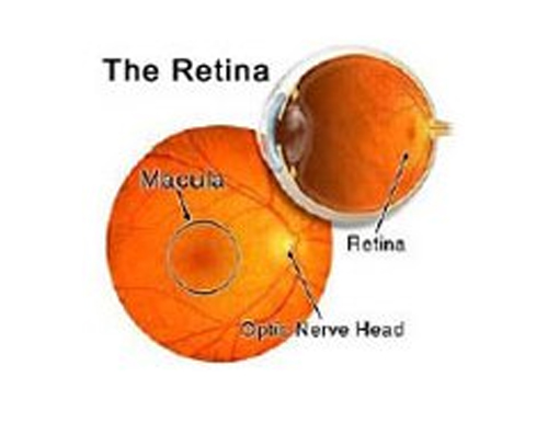
 The Retina is the light-sensitive film in the back of the eye. The image is perceived here and transmitted to the brain by the optic nerve. The vitreous is the clear gel that fills the back of the eye. Diseases of the retina can affect any age.
When to see a doctor
Make an appointment for an eye exam if you notice any changes in your vision. If you develop sudden vision changes, such as double vision or flashes of light, sudden eye pain, or a sudden headache, see your doctor right away.
Common Conditions
Lattice Degeneration
Lattice degeneration is a relatively common condition causing areas of peripheral retinal thinning. Lattice degeneration does not generally present any easily recognizable symptoms. When symptoms are noticed they are usually indicative of a complication (e.g. retinal tear) rather than the lattice condition itself. Most lattice does not require treatment. It is controversial whether associated retinal holes should be prophylactically treated with in-office retinopexy (laser versus cryo), especially in the absence of symptoms. Lattice with retinal breaks (holes and/or tears) remains a leading cause of retinal detachment.
Detached and Torn Retina
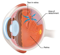 A retinal detachment is a very serious problem that almost always causes blindness unless treated. The appearance of flashes, floating objects, or a gray curtain moving across the field of vision are all indications of a retinal detachment. If any of these occur see an ophthalmologist immediately. As one gets older the vitreous; the clear gel-like substance that fills the inside of the eye tends to shrink slightly and take on a more watery consistency. Sometimes as the vitreous shrinks it exerts enough force on the retina to make it tear. Retinal tears increase the chance of developing a retinal detachment. Fluid vitreous passing through the tear lifts the retina off the back of the eye like wallpaper peeling off a wall. Laser surgery or cryotherapy (freezing) is often used to seal retinal tears and prevent detachment.
Macular Edema
 Macular edema is the swelling of the macula, the small area of the retina responsible for central vision. The edema is caused by fluid leaking from retinal blood vessels. Central vision, used for reading and other close detail work, is affected. Retinal blood vessel obstruction, eye inflammation, diabetes, and age-related macular degeneration have all been associated with macular edema. The macula may also be affected by swelling following cataract extraction, though typically this resolves itself naturally. Eye drops, injections around the eye or laser can be used to treat a macular edema. Recovery depends on the severity of the condition causing the edema.
Myopic Degeneration
People with severe nearsightedness (high myopia) are at greater risk for myopic degeneration. Myopic degeneration commonly occurs during young adulthood and can lead to a gradual decrease in central vision. Vision can decrease more abruptly in a small percentage of patients.
Retinitis Pigmentosa
Retinitis pigmentosa (RP) is the name given to a group of inherited eye diseases that affect the retina. RP causes the degeneration of photoreceptor cells in the retina. As these cells degenerate and die, patients experience progressive vision loss. RP symptoms can vary. In a person with classic or typical RP, night vision and peripheral (or side) vision will be affected initially. Night blindness is one of the earliest and most frequent symptoms of RP. The loss of peripheral vision is often called tunnel vision. Other forms of RP and related diseases include Usher syndrome, Leber’s congenital amaurosis, rod-cone disease, and Bardet-Biedl syndrome, among others.
Recent Research Advances in Retinitis Pigmentosa
Investigations
FFA (FUNDUS Fluorescein Angiography)
Fundus Fluorescien Angiography (FFA)
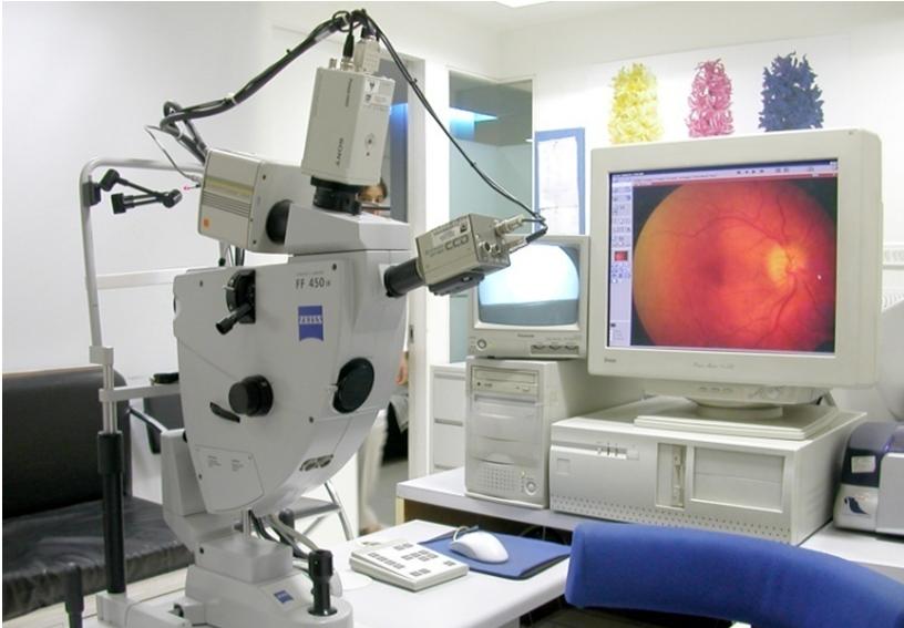 This investigative procedure comprises of injecting a dye -FLUORESCEIN into one of the veins in your arm and either taking rapid serial photographs of its passage within the delicate blood vessels of the eye in the retina and choroid, using a digital Fundus camera or HRA or rarely examining the inside of your eye with an INDIRECT OPHTHALMOSCOPE using appropriate filters.
The information obtained from this test aids your doctor in making a diagnosis and planning treatment especially using different kinds of a laser such as Diode, Argon,532 DF YAG PDT or TTT.
Apart from the needle prick and the light flash of the camera there is no discomfort from this test. If nausea appears to remain calm and take deep breaths which helps in overcoming it. Your usual diet can be taken soon after the procedure. Fluorescein is the highly non-toxic drug. In rare instances, it can produce an allergic reaction, which response rapidly to appropriate medication. Serious life-threatening allergic reactions, though exceptionally rare, can occur. The skin and urine may appear yellow in color for about 36 hours following the test and is not a cause of any concern.
You must be accompanied by an adult attendant during this test.
Instructions to patients
Indocyanine Green Angiography (ICG)
This investigative procedure comprises of injecting a dye -INDOCYANINE GREEN into one of the veins in your arm and either taking rapid serial photographs of its passage within delicate blood vessels of the eye in the retina and choroid, using a digital Fundus camera of HRA using appropriate
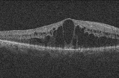
Optical Coherence Tomography
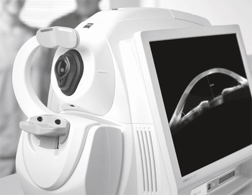 The next generation 3D Optical coherence tomography system. It is a noninvasive, noncontact, transpupillary imaging technology, with ultra-high speed 27,000 axial scans per second which allow 50 times faster data acquisition in practice and high-resolution 5-µm axial and 15-µm transverse resolution in tissue. Fourier domain OCT technology allows the systems advanced clinical protocols to be used for ocular examination with greater resolution and clarity.
The OCT uses light waves to make a map of the retina, anterior chamber, cornea to show up any damaged areas. It uses an optical principle known as low coherence interferometry to scan across the targeted eye structure and generate an image of the same.
The procedure requires you to sit in front of the OCT machine and place your chin on chin rest and forehead touching the head support. You are required to focus at a given target and keep your eye still while the scan is being performed.
The layers within the retina can be differentiated and the retinal thickness can be measured. Also, the change from the previous visit, the effect of treatment can be analyzed.
The optic disc and nerve fiber layer can be assessed and guided progression analysis provides valuable information in the management of Glaucoma
B SCAN
B SCAN is a two-dimensional imaging system which utilizes high-frequency sound waves ranging from 8-10 MHz. It is used for imaging of intraocular structures and giving information about the status of the lens, vitreous, retina, choroid, and sclera.
B-scan ultrasound is most useful when direct visualization of intraocular structures is difficult or impossible in cases like - lid problems (eg, severe edema, partial or total tarsorrhaphy), keratoprosthesis, corneal opacities (eg, scars, severe edema), hyphema, hypopyon, miosis, pupillary membranes, dense cataracts, or vitreous opacities (eg, hemorrhage, inflammatory debris).
In many instances, ultrasound is used for diagnostic purposes even though pathology is clinically visible. Such instances include differentiating iris or ciliary body lesions; ruling out ciliary body detachments; and differentiating intraocular tumors, serious versus hemorrhagic choroidal detachments, rhegmatogenous versus exudative retinal detachments, and disc drusen versus papilledema.
A- scan mode can be used for the measurement of an axial length of the eyeball required for calculation of the lens power before cataract.
Electroretinogram (ERG)
 ERG is the diagnostic procedure to evaluate and confirm diffuse retinal degenerations with the help of mass electric response generated by the retina when subjected to flashes of light.
Multifocal Electroretinogram (MFERG)
 The Multifocal ERG which has been introduced for the first time in Mumbai is used to study local responses from the retina and to determine focal retinal degenerations and lesions.
Electro-Oculogram (EOG)
Is the only means of understanding the measure of retinal function that depends upon the integrity of the retinal pigment epithelium layer of the retina.
Multifocal Visual Evoked Potential (VEP)
Is useful to objectively evaluate visual acuity and aid in perfect diagnosis by topographically mapping brain activity and determining the presence of visual field and visual acuity deficits.
Preferential Hyperacuity Perimeter (PHP)
The most sensitive method in the world to detect the beginning of wet age-related macular degeneration in the most severe form of ARMD and designed to detect and monitor the progression of the disease in a non-invasive manner. The PHP examination is easy to administer and takes just 3-5 minutes providing a comprehensive examination covering 14° of the macular area.
Treatments
Laser Therapy
PhotoDynamic Therapy
Intravitreal injections
Surgeries
The most common indications for a vitrectomy include: